Oral Surgery
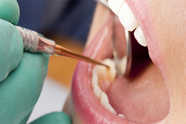
A number of conditions may require oral surgery, including:
Impacted Teeth Wisdom Teeth, otherwise known as third molars, are the last set of teeth to develop. Sometimes these teeth emerge from the gum line and the jaw is large enough to allow room for them, but most of the time, this is not the case. More often, one or more of these third molars fails to emerge in proper alignment or fails to fully emerge through the gum line and becomes entrapped or “impacted” between the jawbone and the gum tissue. Impacted wisdom teeth can result in swelling, pain, and infection of the gum tissue surrounding the wisdom teeth. In addition, impacted wisdom teeth can cause permanent damage to nearby teeth, gums, and bone and can sometimes lead to the formation of cysts or tumors that can destroy sections of the jaw. Therefore, dentists recommend people with impacted wisdom teeth have them surgically removed.
It’s not just wisdom teeth that sometimes become impacted and need to be removed. Other teeth, such as the cuspids and the bicuspids can become impacted and can cause the same types of problems described with impacted wisdom teeth.
Unequal jaw growth In some individuals, the upper and lower jaw fail to grow properly. This can cause difficulty in speaking, eating, swallowing, and breathing. While some of these problems – like improper teeth alignment – can be corrected with braces and other orthodontic appliances, more serious problems require oral surgery to move all or part of the upper jaw, lower jaw, or both into a new position that is more balanced, functional, and healthy.
Improve fit of dentures For first-time denture wearers, oral surgery can be done to correct any irregularities of the jaws prior to creating the dentures to ensure a better fit. Oral surgery can also help long-term denture wearers. Supporting bone often deteriorates over time resulting in dentures that no longer fit properly. In severe cases, an oral surgeon can add a bone graft to areas where little bone remains.
Lesion removal and biopsy Oral surgeons can take a small sample of abnormal growth or tissue and then send it for laboratory testing for identification. Some lesions can be managed medically or can be removed by the oral surgeon.
Facial infections Pain and swelling in the face, neck or jaws may indicate an infection. Infections in this area of the body can sometimes develop into life-threatening emergencies if not treated promptly and effectively. An oral surgeon can assist in diagnosing and treating this problem. Surgical treatment, if needed, may include cutting into and draining the infected area as well as extracting any teeth that might be involved.
Soft-Tissue Grafts Gums can recede for several reasons. They include:
Periodontal disease
Physical trauma (brushing too hard, too often or with a hard brush)
Tooth position
Gum tissue that is naturally thin
If gums recede severely, some of the tooth’s root will be exposed. This can make the tooth sensitive to hot or cold temperatures and more prone to root decay. Some people also don’t like the way it looks.
Soft-tissue grafts are used to add more gum tissue. This can accomplish several things:
Prevent further recession
Cover the exposed root
Stop the sensitivity
Improve the look of the tooth
Some people’s gums are naturally very thin. This increases the likelihood they will recede. In these cases, a soft-tissue graft may be done to prevent problems in the future.
There are three different types of soft-tissue grafts:
Free gingival grafts
Connective-tissue grafts
Pedicle grafts
In a free gingival graft, a small strip of flesh is removed from the roof of the mouth (palate). The tissue, called the “graft,” is then stitched to the gum tissue in the area being treated. A free graft is often used for people who naturally have little gum tissue around their teeth and need to have the gums enlarged.
In a connective-tissue graft, a flap is cut in the roof of the mouth. The flap forms a “trap door” shape. Some tissue from under the flap is removed. The flap is then stitched back down. The tissue that was removed is known as subepithelial connective tissue. This graft tissue is slipped under the gum tissue that surrounds an exposed root. Then it is stitched into place. This is the most commonly used procedure for treating root exposure.
A pedicle graft uses a flap of tissue from the gum of a tooth next to the one with recession. The flap is partially cut away, with one edge still attached. This flap is called a pedicle. It is slid sideways to cover the exposed root and stitched into place. A pedicle graft may be more successful than a free gingival graft. That’s because at least some of the blood vessels that feed the grafted section remain in place. However, a pedicle graft can be done only in certain cases. There must be a tooth next to the one being treated that has enough gum tissue to “share.”
With all of these procedures, many periodontists will use a periodontal dressing. This is a soft material with a texture like modeling clay. It will be used to cover and protect the grafted tissue for the first week or two of healing.
With free grafts and connective-tissue grafts, you will have two wounds. One will be near the tooth; the other will be on the roof of your mouth. Both can create discomfort. The wound on the roof of the mouth is often described as feeling like a significant pizza burn. Prescription pain medicine can help control discomfort after surgery.
Be sure to keep the mouth clean. This means that unaffected teeth will have to be brushed and flossed as usual. Mouth rinses that contain chlorhexidine usually are prescribed to help control plaque during the healing period. Antibiotics may be prescribed, but usually are not.
Typically, there are no long-term side effects after soft-tissue grafting. In some cases, the new gum tissue may need to be reshaped. This procedure is called gingivoplasty. It is used to make the gum look as natural as possible.
Bone Graft (regenerative surgery) A bone graft is a procedure that is used to recreate hard and soft supporting tissues lost due to gum disease.
Regenerative surgery is a treatment for the gum disease called periodontitis. People with periodontitis lose gum coverage and bone support around their teeth. Regenerative surgery regrows these lost tissues.
You need to undergo initial periodontal therapy (scaling and root planing) before your surgical appointment. You also must have a good level of oral hygiene. A local anesthetic is used to numb the area for surgery.
The goal of regenerative surgery is to coax the body into rebuilding the structures that attach a tooth to the jaw, including bone. The dentist will separate the gums from your teeth to gain access to the roots and surrounding bone. The roots will be thoroughly cleaned. The holes (defects) in the bone are filled in with a graft material and covered with a physical barrier.
Bone grafting materials commonly used include bits of a patient’s own bone, cadaver bone, cow bone and synthetic glasses. The patient’s own bone is preferable. The barriers, used to prevent soft tissue from growing into the bony defect, are made from human skin, cow skin or synthetic materials.
The gums are then repositioned over the treated site and secured in place with stitches. During the next six to nine months, your body fills in the periodontal defect with new hard and soft tissue, effectively reattaching the tooth to your jaw.
Pain medications may be prescribed to ease discomfort after surgery.
It is very important for you to keep your mouth as clean as possible while the surgical site is healing. This means you should brush and floss the rest of your mouth normally. If the surgical site is not covered by a periodontal pack, you can use a toothbrush to gently remove plaque from the teeth. Antimicrobial mouth rinses containing chlorhexidine are commonly prescribed after periodontal surgery. Although these rinses do not remove plaque from the teeth, they kill bacteria and help your mouth heal.
You may also have some swelling after surgery. This can be minimized by applying an ice pack to the outside of your face in the treated area. Antibiotics usually are prescribed to prevent an infection and these should be taken as instructed.
After the surgery, you may have some bleeding and swelling. There is a risk that you could develop an infection.
Your gums in the area that was treated are more likely to recede over time. The teeth that were treated may become more sensitive to hot and cold and may develop cavities in the roots.
You will be monitored closely after surgery and examined frequently during the first few months. Always call your dentist if you have excessive bleeding that does not stop.
In addition, call if the surgical area swells up and the pain gets worse one week after the procedure. It may be a sign of developing infection that must be taken care of promptly.
Hemisection is the process of cutting a tooth with two roots in half, creating a front half and a back half. It often is done by a periodontist, but any experienced dentist, endodontist or oral surgeon can do the procedure. Hemisection literally means “dividing in two.”
Because upper molars have three roots, a hemisection cannot be done on an upper molar. However, your dentist could divide an upper molar into three parts (sometimes referred to as trisection), or could remove one or two roots (root resection).
A hemisection is done when decay or bone loss from periodontal disease extends into the area between the two roots, called the bifurcation. The procedure allows your dentist to reach the affected area and provide treatment. Once the roots have been separated, your dentist will evaluate each half of the tooth and its root separately and determine whether they can be restored with crowns. In some situations, this may require crown lenghtening of one or both parts of the tooth and roots. Crown lengthening involves the removal of gum tissue, bone or both to exposure more of a tooth’s structure. In other situations, your dentist may decide that the decay or bone loss is too extensive to allow restoration of one of the halves and roots. In this case, the severely affected half will be removed (resected).
Hemisection is less common than it used to be. In certain instances, guided tissue regeneration can be used to regrow bone in the bifurcation area. In many cases, people choose to have the tooth removed and replaced with an implant. The best option depends on the amount of decay or bone loss affecting the tooth and the probability that it can be successfully restored. You should talk with your dentist about your options.
Ideally, a tooth needing a hemisection should first receive root canal treatment if that has not already been done.
Before hemisection, your dentist also will look at your X-rays and examine your teeth to pinpoint the areas of decay and the extent of periodontal disease.
You will be given local anesthesia for the procedure.
Your dentist often needs to make a small incision in the gum to expose the deeper tooth structures. Once the tooth is exposed, he or she will separate the tooth’s roots. Then your dentist will remove any decay and parts of the tooth that cannot be saved, which could include one of the roots.
The area will then be cleaned by rinsing with sterile saline solution. If necessary, stitches may be used to close the incisions.
The tooth will be covered with a temporary filling or crown, which will later be replaced with a permanent crown.
A hemisection usually takes about an hour from start to finish, but it depends on the amount of decay and periodontal disease present and whether other procedures are necessary.
You probably will feel some discomfort and swelling and have light bleeding for the next 24 to 48 hours. Avoid chewing with the tooth until the stitches are removed. Your dentist may prescribe an antimicrobial mouth rinse.
After 7 to 10 days, you will visit the dentist to have stitches removed. Your dentist also will check to see how your gums are healing.
After a few months, the tooth should be healed enough for your dentist to place a crown. You may receive one crown that fits over both halves, leaving a space between the two roots to allow easy access for oral hygiene. Alternatively, the two halves of the tooth can be restored as individual teeth by placing two separate crowns, one on each root. It is even possible to use individual tooth roots to help support bridges. For example, if you had two adjacent teeth hemisected and the two middle halves removed, your dentist could put a bridge in the space and support it with the two remaining halves.
A tooth that has been hemisected may be more difficult to keep clean. Also, the tooth will not be as strong as it was, especially if one of the roots has been removed, and is therefore more susceptible to fracture.
If the tooth develops further complications, it may need to be extracted.
If you are concerned about the level of pain, swelling or bleeding in the days after the procedure, contact your dentist.
Apicoectomy Your teeth are held in place by roots that extend into your jawbone. Front teeth usually have one root. Other teeth, such as your premolars and molars, have two or more roots. The tip or end of each root is called the apex. Nerves and blood vessels enter the tooth through the apex. They travel through a canal inside the root, and into the pulp chamber. This chamber is inside the crown (the part of the tooth you can see in the mouth).
During root canal treatment, the canals are cleaned. Inflamed or infected tissue is removed. An apicoectomy may be needed when an infection develops or won’t go away after root canal treament or retreatment.
Root canals are very complex, with many small branches off the main canal. Sometimes, even after root canal treatment, infected debris can remain in these branches. This can possibly prevent healing or cause re-infection later. In an apicoectomy, the root tip, or apex, is removed along with the infected tissue. A filling is then placed to seal the end of the root.
If a root canal procedure has been done in the past and it becomes infected again, it’s often because of a problem near the apex of the root. In many cases, a second root canal treatment is considered before an apicoectomy. With advances in technology, dentists often can detect other canals that were not adequately treated. In this case, they may be able to clear up the infection by doing a second root canal procedure. This will avoid the need for an apicoectomy.
Your dentist can do an apicoectomy to fix the problem so the tooth doesn’t need to be extracted. An apicoectomy is done only after a tooth has had at least one root canal procedure and retreatment has not been successful or is not possible.
For example, retreatment is often not a good option when a tooth has a crown or is part of a bridge. Retreatment of the root canal would require cutting through the crown or bridge. That might destroy or weaken the crown or bridge. An apicoectomy is often considered in a situation like this.
An apicoectomy is not the same as a root resection. In a root resection, an entire root is removed, rather than just the tip.
Before the surgery, your dentist may take more X-rays of the tooth and surrounding bone. You may be given an antimicrobial mouth rinse, a medicine to reduce inflammation, and/or antibiotics.
Your dentist also will review your medical history. Depending on other existing medical conditions, your dentist may consult with your physician before the procedure.
Your gum will be cut and lifted away from the tooth to gain access to the root. The infected tissue will be removed along with the last few millimeters of the root tip. The endodontist will use a dye to highlight cracks and breaks in the tooth. If the tooth has large cracks or breaks, it may have to be extracted. In this case, the apicoectomy will not continue.
To complete the apicoectomy, the end of the tooth’s canal will be cleaned and sealed. The cleaning usually is done under a special microscope using ultrasonic instruments. The light and magnification allow the endodontist to see the area clearly. This increases the chance that the procedure will succeed. Then an X-ray will be taken of the area before the tissue is stitched back in place.
Most apicoectomies take 30 to 90 minutes. The length will depend on the location of the tooth and the complexity of the root structure. Procedures on front teeth are generally the shortest. Those on lower molars generally take the longest.
You will be told which medicines to take and what you can eat or drink. You should apply ice to the area for 10 to 12 hours after the surgery, and rest during that time.
The area may bruise and swell. It may be more swollen the second day after the procedure than the first day. Any pain usually can be controlled with over-the-counter nonsteroidal anti-inflammatory drugs (NSAIDs), such as ibuprofem (Advil, Motrin and others). In some cases, you may be given a prescription medicine.
To allow for healing, you should avoid brushing the area or rinsing vigorously. Also, don’t smoke or eat crunchy or hard foods. Do not lift your lip to examine the area. This can loosen the stitches and disrupt formation of a blood clot that is needed for healing.
You may have some numbness in the area for days or weeks after the surgery. Tell your dentist about any numbness. The numbness usually goes away with time.
Your stitches will be removed 7 days after the procedure. All soreness and swelling are usually gone within 14 days.
Even though an apicoectomy is considered surgery, many people say that recovering from it is easier than recovering from the original root canal treatment.
At the consultation the risks of the procedure will be reviewed. The main risk is that the surgery may not work and the tooth may need to be extracted.
Depending on where the tooth is located, there may be other risks. If the tooth is in the back of your upper jaw, the infection can involve your sinuses. Your dentist may suggest antibiotics and decongestants.
An apicoectomy is usually a permanent solution. It should last for the life of the tooth.
If you’re having any pain or swelling from a tooth that has had root-canal treatment, contact your dentist. Sometimes after a root canal a pimple develops near the tooth. This pimple will often go away and then come back. This is called a fistula. The fistula is a sign that there is an infection and your body is draining it out through the pimple. There is usually no pain in this situation.
If you have symptoms, such as pain or a fistula, contact your dentist. He or she will take X-rays and do an exam. If your dentist feels you need an apicoectomy, you will need to set up an appointment for a consultation.